The Journal29 August 202612 min read
A man who could only say “Tan”
How one lost word mapped the brain — and how the map went out of date
The man they called “Tan” at the Bicêtre hospice in Paris was named Louis Victor Leborgne. When Paul Broca examined him in 1861 he was 51 years old and had lived in the institution for decades. He understood a good deal, answered with gestures, showed numbers with his fingers — and yet produced almost nothing but that one syllable. Latterly a right-sided paralysis was added. A few days after Broca’s examination he died, on 17 April 1861. The autopsy revealed extensive damage in the left frontal lobe.
Broca presented the brain the very next day. The case seemed to settle one of the great questions of the age: does the brain work as an undivided whole, or do particular regions have particular tasks? A man who loses articulate speech and carries a lesion at the front left is a strong argument for the second answer. Out of this specimen came a map, out of the map a point in the textbooks — and out of the man a syllable.
Il comprenait tout ce qu’on lui disait; il avait même l’oreille très fine; mais, quelle que fût la question qu’on lui adressât, il répondait toujours: tan, tan, en y joignant des gestes très variés au moyen desquels il réussissait à exprimer la plupart de ses idées.
The hall, the question, the specimen
Broca was not the first to suspect that speech lay in the frontal lobe. Jean-Baptiste Bouillaud had collected cases. His son-in-law Ernest Auburtin argued before the Paris Anthropological Society in early April 1861 that lesions of the anterior lobes could abolish speech without destroying the other mental faculties. He challenged his opponents, in effect, to produce a case that refuted it. Broca was in the audience. A few days later Leborgne was on his ward.
That does not diminish the observation, but it changes the dramaturgy. Broca did not look at a brain without preconceptions and find language in it. A scientific community had already prepared the hypothesis, the counter-argument and the criterion of proof. The case became famous because it fitted an open problem and because Broca demonstrated the specimen convincingly.
Someone else had the finding before him. In the 1830s Marc Dax had come to the conviction that loss of speech was connected with the left half of the brain; he drew on some eighty cases from the literature and from his own practice. Whether he actually presented them at Montpellier in 1836 is disputed among historians; the work was not published in his lifetime. His son Gustave made good the omission in 1865, four years after Broca’s presentation, and claimed priority.
The dispute shows how many different things “discovery” can mean: being the first to voice an idea, gathering data, publishing, persuading a professional community, founding a research programme. In none of these roles was Broca the first. He merely combined them more effectively than anyone else.
Broca tested whether Leborgne understood instructions, whether he could indicate numbers with his fingers and carry out movements. One distinction was decisive: tongue and larynx were not paralysed, the speech musculature not mechanically unusable. What had been lost was the capacity to organise meaningful sequences of sounds.
The examination lasted only a few days, because the patient was gravely ill. That heightened the drama and limited the depth of testing. Naming, repetition, reading, writing, sentence comprehension, non-verbal problem solving — the examinations we now take for granted had not yet been standardised.
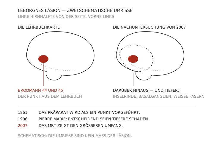
The finding, too, was more complex than it appeared. On the surface the lesion in the left frontal lobe stood out; beneath it lay extensive damage to the insular cortex, the basal ganglia and white-matter connections. The disease had probably developed over years. To link a single surface with a single symptom was heuristically fruitful — and anatomically cruder than the later textbook illustrations suggest.
Three decades in the hospice
Leborgne was born in 1809 and came to Bicêtre at about thirty, after largely losing the ability to speak. The scant record describes him as a former artisan; much else remains uncertain. He lived some 21 years with the speech disorder, the last of them with increasing paralysis. When Broca saw him the illness was therefore not recent but the end of a long neurological course.
Bicêtre was at once a hospital, a hospice and a place of confinement for men at the margins of society. Records of that kind preserve symptoms, beds and medical decisions, hardly any friendships or self-interpretations. Leborgne’s perspective is obscured twice over: by the logic of the archive and by the aphasia that made a detailed counter-narrative difficult for him.
Contemporary reports describe him answering with gestures and swearing when frustrated. That proves neither a particular character nor complete comprehension of speech. It shows that communication does not end at grammatically complete sentences. Facial expression, gaze, rhythm, pointing and the shared situation carry meaning. Anyone who counts only the missing words overlooks how a person acts nonetheless. “Tan” is therefore not merely a clinical sign but the mark of a gradient: between a patient who could barely be quoted and doctors whose papers are still read today.
| Detail | Value |
|---|---|
| Birth of Louis Victor Leborgne | 1809 |
| Loss of speech from about | his thirtieth year |
| Duration of the speech disorder | about 21 years |
| Examination by Broca and death | April 1861 |
| Presentation of the brain | the day after his death |
| Extent of the lesion on MRI | beyond Brodmann 44 and 45 (Dronkers et al., 2007) |
| First major contradiction | Pierre Marie, 1906 |
From centre to network
Broca’s case established the lesion-symptom method: where a circumscribed injury coincides with a specific deficit, one can infer the function of a system. In 1874 Carl Wernicke described patients with fluent but barely intelligible speech and impaired comprehension. He linked this pattern to left posterior temporal regions and proposed connections between sensory and motor images of speech. Later diagrams turned this into two boxes with a cable between them.
The model was productive because it predicted different patterns of error, among them a conduction aphasia when the connection is disturbed. It is also too simple. Language uses dorsal and ventral pathways, temporal, frontal and parietal regions as well as subcortical structures. Meaning, sound structure and the preparation of action are not processed one after another at two stations. Historical models should therefore be neither mocked nor taken for settled anatomy. Part of their value lies in the more precise questions their errors made possible later.
The objection came early. In 1906 the neurologist Pierre Marie attacked the classical doctrine: the region named after Broca did not by itself explain motor aphasia, he argued; what mattered were deeper and more far-reaching lesions. Later research proved neither side entirely right. Left frontal regions are causally important; but persistent severe disorders of production usually arise from larger network damage.
Sign languages also corrected the equation of language with sound. Left-hemisphere networks process grammatical and lexical structures even when hands rather than voice express them. Leborgne’s problem therefore concerned not merely the muscles of articulation but part of the capacity to translate structured symbols into action. The region named after Broca remains important for language production and sequential processing. It is not a dictionary, and it is not the sole “seat” of language either.
Broca’s first formulation emphasised the frontal lobe; the left side moved to the centre only after further cases. In the majority of right-handed people a large part of the organisation of language lies on the left. Many left-handers also show left dominance, some a stronger right-sided or bilateral involvement. Lateralisation is a statistical tendency, not a fixed label.
Development plays a part. If one hemisphere is damaged early, other networks can take over functions to a greater degree. After a stroke in adulthood reorganisation is likewise possible, but constrained by the existing specialisation and the extent of the lesion. Plasticity does not mean that any region can take on any task at any time.
The popular left-right myths — left logical, right creative — cannot be derived from Broca’s finding. Complex thought uses both hemispheres. The asymmetry is real, but it concerns concrete processing contributions and not two opposed personality types. From a lateralisation of language follows neither character nor aptitude for a profession.
The preserved brain under modern scanners
Leborgne’s brain was not cut into sections but kept as a specimen. It could therefore be examined more than a century later with high-resolution magnetic resonance imaging. In 2007 Dronkers and colleagues showed that the damage went far beyond the traditionally marked Brodmann areas 44 and 45. Broca’s second famous patient, Lelong, also had a more complex lesion pattern than the textbooks suggested.
The re-examination matters methodologically. A historical name can feign an anatomical certainty that the historical object itself does not support. “Broca’s area” was defined from many later findings; it is not simply the site in Leborgne’s brain. Conversely, the larger lesion does not prove that the left frontal cortex is unimportant. It shows only that a single case could not demonstrate its necessity in isolation.
Today lesion-symptom maps compare many patients statistically. But vascular territories produce typical combinations of lesions, and neighbouring regions are therefore hard to separate. Functional imaging, for its part, shows where a blood-flow signal changes during a task; activity may be necessary, supporting, compensatory or merely a consequence of other processing. Direct electrical stimulation disrupts a region temporarily but is used only where there is a clinical reason. Transcranial magnetic stimulation is less invasive and does not reach every structure precisely.
A dependable statement therefore emerges from convergence. When imaging, lesion, stimulation and time course all point to the same mechanism, confidence grows. Broca’s methodological legacy is not a point on the map but the idea of relating behaviour and anatomical disturbance to one another systematically — and of testing that relation afresh with every new method.
Aphasia in daily life, Broca in retrospect
Classic Broca’s aphasia is described in terms of effortful, non-fluent speech, abbreviated grammar and relatively better comprehension. In practice the profiles vary widely. Some patients cannot find words, others founder on complex syntax, on planning sounds, or on repetition. Comprehension is by no means always intact for grammatically demanding sentences.
The distinctions are more than terminology. Apraxia of speech means a disorder of the motor planning of speech movements, dysarthria the neuromuscular execution; both can occur alongside an aphasia and are not the same thing as one. Leborgne’s historical record does not allow these levels to be cleanly separated; modern assessment examines types of error, speech melody, mouth movements and comprehension separately. Therapy has to know whether someone cannot find a word, cannot plan its sequence of sounds, or cannot control the muscles. And for relatives it makes a difference whether a short answer arises from failing comprehension, laborious production or exhaustion.
The course of the condition forbids quick judgement. In the first days after brain damage, swelling, reduced blood flow and general illness intensify the symptoms. Spontaneous recovery and therapy change the profile over weeks and months; a single test is therefore no final verdict on someone’s capacity to communicate. Some people regain a great deal of spoken language, others develop effective strategies despite a persisting aphasia. With multilingual people there is the added fact that languages use overlapping networks and are nevertheless differently vulnerable: age of acquisition, frequency of use, emotional significance and structural similarity all help determine which remains more accessible after a lesion. Testing in only one language mistakes low familiarity with the test for neurological loss.
Disability also arises between people. An impatient interlocutor, a form with no support, a hectic hospital all enlarge the problem. Time, pictures, yes-no options and respectful follow-up questions make visible abilities that stay hidden in rapid conversation. For fluency carries a social halo: those who speak quickly and elegantly seem more competent, those who falter are underestimated. Intelligence tests, too, judge aphasic people unfairly when their tasks depend needlessly on language.
This is not merely a matter of courtesy. Institutions tie participation to linguistic speed — in the consulting room, at the public office, in court. Anyone who equates expression with the capacity for judgement strips people of rights precisely where they need more time and support. Leborgne’s preserved responses already suggested in 1861 that articulate speech and general thought are not the same. A person can know who they are looking for and not find the name; they can feel an injustice and barely be able to formulate the grounds for it.
Paul Broca was a surgeon, an anatomist, a founder of scientific societies and a politically engaged republican. He developed measuring instruments and at the same time upheld hierarchies between the sexes and between population groups that his skull measurements were supposed to support. His figures were not free of social assumptions about who counted as more highly developed.
It would be too easy to discard the language finding because of these errors. It would be equally wrong to read scientific achievement as proof of moral foresight. The same person can make a precise clinical observation and, in other areas, dress up prejudice as method. Science does not automatically protect against ideology; it needs criticism of selection, measurement and interpretation.
Posthumous fame, moreover, follows the Matthew effect: well-known researchers and memorable names accumulate further credit, while patients, forerunners and collaborators fade. “Broca’s area” is a practical term. It inscribes a social history into anatomy.
What remains
Four degrees of certainty order the case. It is certain that Leborgne had a severe disorder of spoken language and a large left-hemisphere lesion. It is strongly supported that left frontal networks and those connected with them are causally important for language production. It is less certain which sub-region produced which part of his profile. It remains unanswerable how he himself experienced the decades of restriction. This gradation preserves the value of the insight without declaring the patient a perfect natural experiment: diseases do not respect textbook boundaries, and a life supplies no control condition.
Anyone who hears today that language sits in Broca’s area has a sentence from 1861 in their ear, one that the findings have worn down ever since. The case remains a large one precisely when it is told smaller: it proved no seat but established a method that infers functional organisation from selective losses. The following 165 years consisted in refining that first map. What is more useful in conversation than any map can be said in a single sentence: a person who cannot find a word has not lost the thought.
Sources, and why they are here
Broca, P. (1861). Remarques sur le siège de la faculté du langage articulé, suivies d’une observation d’aphémie. Bulletins de la Société Anatomique, 6, 330–357.
The original report on Leborgne — source of the quotation and of every detail on the examination and autopsy of 1861.
Dronkers, N. F., et al. (2007). Paul Broca’s historic cases: high resolution MR imaging of the brains of Leborgne and Lelong. Brain, 130(5), 1432–1441.
The MRI re-examination of the preserved specimens; it establishes that the damage reached far beyond areas 44 and 45.
Cubelli, R., & De Bastiani, P. (2011). 150 years after Leborgne. Cortex, 47(2), 146–147.
Places the case 150 years on and separates what Broca showed from what was attributed to him.
Tremblay, P., & Dick, A. S. (2016). Broca and Wernicke are dead, or moving past the classic model of language neurobiology. Brain and Language, 162, 60–71.
Explains why the two-centre model no longer carries the present evidence — the basis of the section on networks.